Hepatic Toxicology
Here's the location and overview of the liver. Go to
http://www.innerbody.com/ , click on block, Guide to the Human Body, and then Digestive System, find the liver and the gallbladder, a little storage tank for bile laying next to the liver,
which is fed by two main ducts from the liver. You should read the sidebar on the right about liver and gallbladder.
The liver receives virtually all the blood flow from the GI tract and processes
the blood. Nutrients and other proteins and chemicals are added to the blood
or taken from the blood in the liver. It is an amazing organ in a blob-like
structure. Because the blood flowing into the liver from the GI tract is all
venous blood, it is oxygen poor. The vein that carries this oxygen poor blood
into the liver is called the portal vein. After blood leaves the liver, it flows
in the hepatic vein towards the heart. The liver needs oxygen for all the work
it must do. Oxygen is supplied from oxygen-rich blood from the hepatic artery.
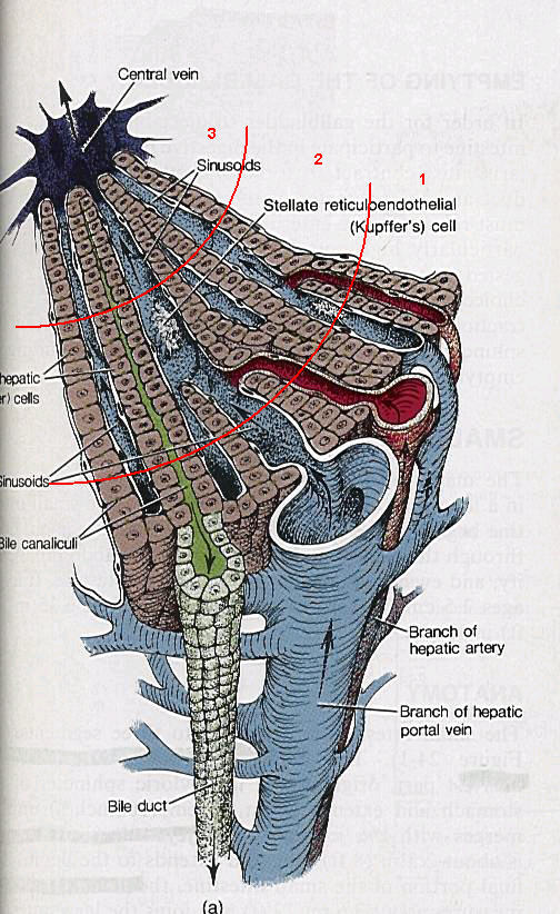 |
Here is a neat diagram of liver function. The predominant cell type is
the hepatocyte, shown in brown. Blood from the GI tract flows in via the
portal vein and flows out through the central vein. Oxygen-rich blood
from the hepatic artery mixes with the venous blood from the portal vein.
Concurrently bile from the hepatocytes flows towards the bile duct (it
looks to me like a back alley). The grouping of the portal vein, hepatic
artery and bile duct is called the "portal triad." The street
the blood flows through is called a sinusoid. The funny white cells have
two names, I'll give you a third, they are macrophages. They scarf any
bacteria that make it in from the GI track and also gobble things like
wounded or worn-out red blood cells. Also shown are three zones. Zone
1 has the most oxygenated blood and zone 3 the least oxygenated. For simplicity
I showed you one set of influent vessels and one set of effluent vessels.
Because there are lots of triads and central vein branches, you could
describe an aerial view of a liver section in a honeycomb arrangement
of hexagons with a triad in each vertex and central vein in the center
of each hexagon, that is called the lobule view. Figure 11.3 of your book
shows the acinus view, with flow from one triad flowing towards four central
veins (two are shown).
Picture from Tortora and Anagnostakos, 5th edition, copyrighted
|
The next site has some neat
photographs of liver micro anatomy, but you only need to look at the photos
I mention. Photos 1 and 2 show the basic lobular arrangement. Photo three shows
a triad. Photo 7, at a much higher magnification, shows the "space of disse." But if that site does not come up, try this one
http://en.wikipedia.org/wiki/Perisinusoidal_space The sinusoids are blood vessels, lined with endothelium. But it is a special
type of endothelium, with windows that let the blood circulate out of the sinusoid
to get closer to the hepatocyte (kind of the opposite of a blood-brain barrier).
The wall of the hepatocyte has little hair-like projections, microvilli, that
vastly increase the surface area of the cell in contact with the blood.(It doesn't
say, but my guess is the width of the photograph is 0.1 micron.)
The liver stores nutrients, sugar and fats. It also produces blood proteins.
We'll show some chemical damage to the liver in the section on alcohol. Here
I want to mention the phenomenon of cholestasis where damage to the liver
results in the obstruction of bile flow. Bilirubin, a breakdown product of red
blood cells, is normally excreted via the bile. It is responsible for the brown
coloration of feces. When bile flow is blocked, bilirubin builds up in the blood
and causes the yellowing of the skin known as jaundice. The feces may turn very
light colored.
We will mention the specific action of chemicals on the liver when we discus
agents, but most toxic agents damage the liver.
The liver regenerates, that is, if cells are damaged, new cells will form.
The new cells are formed in zone 1, where there is the most oxygen. They then
migrate down the sinusoid toward zone 3. Microscopic analysis has shown that
certain chemical cause their lesions in certain zones, perhaps indicating differing
metabolic process associated with different oxygen tensions in the zones.
The liver is responsible for the processing of fat. You learned from that pdf
file from SciAm, the role of lipids in atherosclerosis Very briefly, LDL supplies
fat to the lesion, and HDL removes it. LDL = bad; HDL = good. Beyond that, there
is an enormous amount of information and research involving fat transport, the
liver, and its effect on atherosclerosis. Most chemicals that can disrupt the
liver will disrupt lipid transport.
Module 08 Index