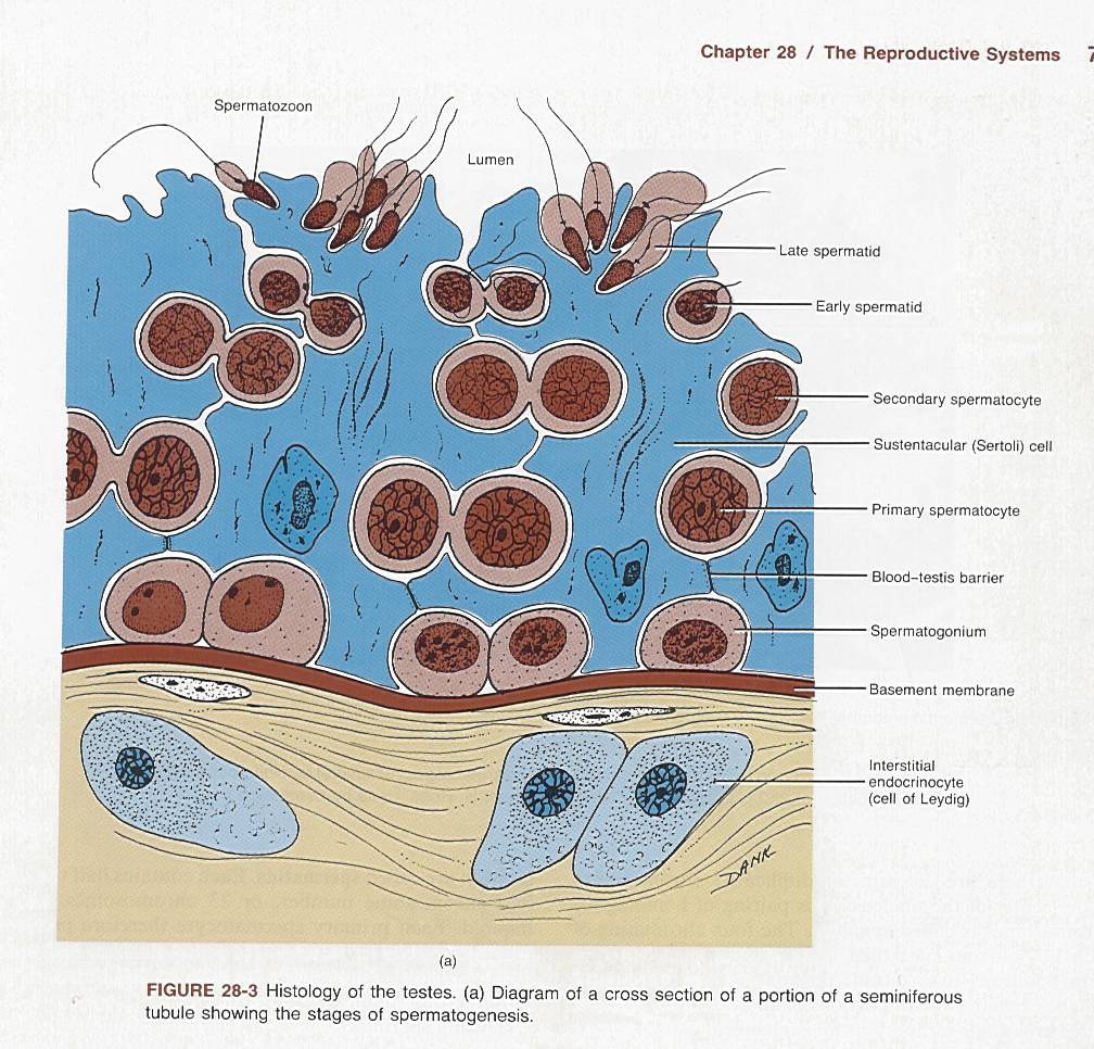
Male germ tissue
Here's a diagram of a cross section through a seminiferous tubule. The "lumen" is the channel of a tubular organ, i.e., water flows through the lumen of a pipe. Shown are stages in the maturation of spermatozoon from the germ tissue, the spermatogonium, a diploid cell (two sets of chromosomes) to the spermatozoa, a haploid cell (one set of chromosomes). The basement membrane is connective material secreted by epithelial cells and connective tissue cells. Above the basement membrane are cells that support the maturation process, called Sertoli cells (aka sustentacular cells). Below the basement membrane are the Lydig cells. The Lydig cells are endocrine cells that secrete testosterone. Between the Lydig cells are shown some connective tissue cells or myoid cells (smooth muscle tissue). Not shown well, because it is not distinct, is the blood-testis barrier. Blood is supplied to the region below the basement membrane by capillaries. In order for a chemical to transfer between blood and spermatozoon, the chemical must traverse the capillary lining (endothelium, the cells that make up the capillary), the connective tissue and myoid cells, the the basement membrane, the Sertoli cells, to reach the spermatozoa.
Now the Lydig cells are stimulated to produce testosterone by the hormone LH (luteinizing hormone), and the Sertoli cells and germ cells (spermatogonium) are stimulated by a hormone called FSH (follicle stimulating hormone). The endocrine system is characterized by feedback control circuits. Below is shown a schematic of the control of secretion of testosterone.
Lets start at the top. The hypothalamus is part of the CNS, i.e., nerve tissue. Hanging directly below the hypothalamus is the pituitary gland. The pituitary is the body's master gland, it controls the function of other glands. The pituitary has two parts, one part secretes hormones directly from a type of nerve tissue, and one part secretes other hormones from gland (epithelial ) tissue. FSH and LH are secreted by the glandular part of the pituitary. (FSH and LH are called gonadotropic hormones.) Above the number one indicates that the hypothalamus secretes a hormone GnRF (Gonadotropin Releasing Factor) into the blood supply of the pituitary. That stimulates the pituitary to release FSH and LH, shown as step 2. The FSH enters the blood and is circulated to the seminiferous tubules of the testes, step 3, where it stimulated spermatogenesis and the supporting cells, the Sertoli cells step 4. In a parallel track, LH stimulates the Leydig cells to secrete testosterone. Step 5 indicates the circulating testosterone stimulates muscle development, and male secondary sexual characteristics (including baldness). The circulating testosterone does several things. It directly inhibits the secretion of LH by the pituitary (step 7) and it also inhibits the secretion of GnRF from the hypothalamus (step 6). When these inhibitions lead to a decrease in the circulating testosterone, this stimulates the hypothalamus to increase the secretion of GnRF. A similar feedback process exists between the hormone inhibin and FSH.
Understanding feedback control, you can now see how anabolic steroids, the illegal drugs some athletes take to increase muscle mass, can make them sterile. Taking large quantities of the drug, which is testosterone or a testosterone analog, inhibits the secretion of GnRF the same as endogenous testosterone would. The lack of GnRF leads to a decrease in the production of LH and FSH, which shuts down production by the Sertoli cells which support the spermatogenesis. Long term high dosage use of the these drugs can lead to atrophy of the seminiferous tubules and perhaps permanent damage.
Spermatozoa have a life of about 90 days and are continuously replaced.
The messenger GnRF is a simple polypeptide of 10 amino acid. LH, FSH are relatively large proteins consisting of about 200 amino acids and some sugar groups. Testosterone and the estrogen are lipophilic hormones made from cholesterol. They travel about in the blood in carrier proteins that are usually specific to the hormone. The hormones are not covalently bound to the carrier and some hormone will travel in the blood unbound from the carrier in the blood.
Female tissue.
The female system is similar in basic chemicals, but is cyclically controlled by the hypothalamus and controlled during pregnancy by hormones secreted by the placenta. A very important point about the female reproductive system is that all the eggs are produced while the female is in utero. That is, a female is born with all the oocytes she will have. The implications from this is that the oocytes are subject to chemical insults for many years, while the sperm are only available for 90 days.